

On the 2nd to 5th April 2019, a team of Insigneo researchers made up of Dr Bryant Roberts, Dr Vee San Cheong, Dr Dharshini Sreenivasan, Mr Liam Boyle and led by MultiSim researcher Dr Enrico Dall’Ara attended the Diamond Light Source I13 X-ray Imaging Lab in the Harwell Science and Innovation Campus, Oxfordshire.
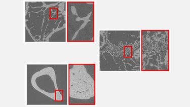
Here the team acquired 3D images of the mouse tibia cortical and trabecular bone using pink light, which at the I13 beamline has flux over a million times higher than that of a laboratory X-ray source. This enabled the very rapid imaging of our specimens at a higher resolution (with 1.6 µm voxel size) and with a higher signal to noise ratio than is achievable with desktop micro-computed tomography (microCT).
The data generated will be used to investigate the effects of pharmacological treatment and mechanical stimuli on the mouse bone tissue. This includes studying the individual and combined effects of bone anabolic treatments, including parathyroid hormone and mechanical loading, in a mouse model with oestrogen-deficiency related bone loss. This research will extend findings from two recent longitudinal microCT imaging studies within MULTISIM to examine variations in the osteocyte lacunae distribution and vascular canal morphology; nanostructures that could not be resolved using standard Desktop microCT systems in a reasonable time.
Commented Dr Bryant Roberts: “The opportunity to contribute in the formulation and writing of a research proposal for the Diamond Light Beamtime was an invaluable experience in my growth as an early career researcher. I look forward in future to realising the exciting outputs from this work as we reveal new insights into treatment effects at the different dimensional scales of bone.”
Team leader, Dr Enrico Dall’Ara commented: “It is every time great to perform experiments at the Diamond Light Source! This data will allow us to better understand the effect of interventions for the musculoskeletal system and to build our computational models to reduce, refine and replace the usage of animals in research.”
Beamtime on the Diamond Manchester Imaging Beamline (I13-2) was awarded to Dr Enrico Dall’Ara and Dr Bryant Roberts for proposal No: MG21628 “Assessing the individual and combined effects of parathyroid hormone (1-34) and mechanical loading on the micro- and nano-structural properties of murine cortical bone”. This study was partially funded by EPSRC MultiSim Grant: EP/K03877X/1, UK National Centre for Replacement, Refinement and Reduction of Animals in Research Grant: NC/R001073/1 and by Diamond Light Source.