X-ray Crystallography
The X-ray crystallography laboratory in the School of Mathematical and Physical Sciences is both a user and service facility. We are equipped with three single crystal and two powder diffractometers.

Services
The X-ray crystallography service offers a complete service for the collection, structure solution and analysis of samples as well as producing publication ready graphical outputs and reports for small molecules.
Single crystal data collections can be undertaken over a wide range of temperatures from 28 K to 500 K.
We can also offer advice on crystallisation techniques to get the best quality samples for your research.
Lab and instruments
Single Crystal
- Bruker AXS Venture
-
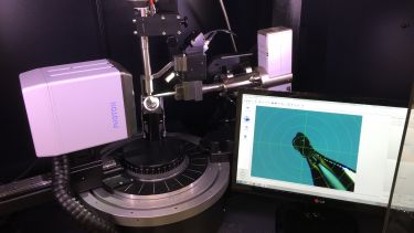
Our newest single crystal diffractometer is the D8 Venture. The Venture is equipped with a high brightness Iμs microfocus Cu source and large Photon III C14 X-Ray detector collecting in shutterless mode. The more intense x-ray beam and increased sensitivity of the detector makes it ideal for small/weakly diffracting crystals and determination of absolute configuration of small organic molecules with no atoms heavier than oxygen and crystals with large unit cells. An Oxford 700 Cryostream allows for data collection over a temperature range of 80–500 K
At-a-glance features:
- High brightness Iμs microfocus Cu source ~150 μm beam width
- Photon III C14 detector
- Three circle goniometer
- Oxford cryostream 700 plus (80–500 K)
- Bruker AXS Smart
-
The SMART is our 3-circle diffractometer equipped with a Mo sealed source, MONOCAP collimator for improved intensity, Oxford 700 Cryostream (80-400 ˚K) and an Apex II CCD detector. SMART is extensively used for routine data collection.
At-a-glance features:
- Sealed Mo source - ~500 μm beam width MONOCAP optics to enhance beam intensity
- Apex II CCD detector
- Three circle goniometer
- Oxford cryostream 700 (80–400˚ K)
- Bruker Nonius - Kappa
-
The KAPPA is our 4-circle diffractometer with a Mo Sealed source and Apex II CCD detector which is used for routine data collection while equipped with an Oxford 700 Cryostream (80-400 ˚K) and best utilised for slightly smaller crystals than would be collected on the SMART. In addition, it also equipped with an Oxford N-HeliX low temperature cooling system allowing collection temperatures as low as 28 ˚K for Charge Density Studies.
At-a-glance features:
- Sealed Mo source ~350 μm beam width MIRACOL optics to enhance beam intensity
- Apex II CCD detector
- Four circle goniometer
- Oxford cryostream 700 (80–400˚ K) Or N-HeliX Nitrogen/Helium Cooler (28-300˚ K)
Powder
- Bruker AXS - Advance
-
The D8 Advance is the newest powder diffractometer in our department. The advance runs a sealed Cu source and LynxEye detector for high quality in house data collections. The Advance may be run with the Flat Plate stage or Capillary stage installed for versatility. A Oxford cryostream can be used in capillary mode to run data collections at a range of (80–500˚ K).
At-a-glance features:
- Sealed Cu source
- Capillary Sample Stage or Flat Plate Sample Stage
- Focusing Göbel Mirror in Capillary Mode
- Variable slit motor in Flat Plate
- LYNXEye detector
- Oxford cryostream 700 plus (80–500˚ K) in Capillary mode
Specialist features by request:
- Anton Paar High Pressure Powder Cell – in collaboration with Dr Marco Conte
- Bruker AXS - D8 Series One
-
Our Series One D8 uses a Cu sealed X-ray source with a scintilating detector. Whilst our Advance has largely super-seeded the technology of the Series One, the machine is routinely used for polymer analysis.
At-a-glance features:
- Sealed Cu Source
- Improved resolution with soller slits
- Scintillation detector
- Flat plate stage
- Heated stage
Other equipment
Polarising microscope
Our polarising microscope has a rotating plate which allows us to conveniently inspect crystal samples. This is to ensure they are of appropriate quality to be used.
A scale embedded in one of the eyepiece lens allows us to roughly measure the crystal size so the best suited mounting tip can be used and the most appropriate diffractometer is used.
Video Microscope:
Our polarising video microscope is connected to a computer allowing the projection of the microscope to aid teaching and training of new users.
Images and videos of samples may be recorded for inclusion in reports if required.
Contact
The X-ray crystallography suite is located in H03 in the top of the North Wing of the Dainton Building.
Email: Dr Craig C. Robertson, craig.robertson@sheffield.ac.uk.
