
Saving Young Lungs - Zoe's Story
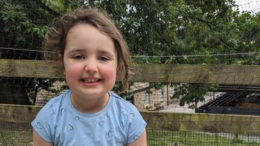
Zoe is 5 years old and loves cycling, swimming and playing board games. And thanks to MRI imaging technology developed by the Insigneo Institute at the University of Sheffield, she’s able to continue doing the things she enjoys, despite suffering from a serious lung condition.

I knew that something was wrong when Zoe developed a cold that just kept getting worse. And after hours in A&E, Zoe was diagnosed with Adenovirus Pneumonia. With low oxygen levels and breathing struggles, we had to stay in hospital for 5 days. I knew that it was going to be a slow road to recovery.
But weeks passed and Zoe kept getting worse. We spent weeks in and out of A&E and the GP surgery, always being sent home without answers. I knew that something was wrong, so I pursued a diagnosis for Zoe. We went through countless X-rays and CT scans, bloods and even more tests.
Eventually, Zoe was diagnosed with bronchiectasis, a complication following pneumonia. For Zoe, breathing can be difficult.
Her lungs are inflamed, producing mucus that acts as a breeding ground for bacteria. The bacteria creates more damage which leads to inflammation, creating an endless cycle.
Zoe was put on IV antibiotics at just 4 years old. And with Zoe potentially needing this treatment on a regular basis for the rest of her life, we were faced with the heartwrenching decision as to whether Zoe should undergo invasive surgery to have a portacath put in. Zoe is an active child, so this course of treatment would’ve had a huge impact on her day to day life. I wanted to be really sure that this was going to be worthwhile.
But thanks to a new type of MRI imaging being trialled in Sheffield, doctors were able to see inside Zoe’s lungs.
They saw that she was not improving with the antibiotics, and so the portacath would’ve been unnecessary, it meant we were able to explore other treatment options, and Zoe was saved from having to undergo invasive surgery.
I'm so grateful that we've had the opportunity to use the MRI imaging. And I think it should be available to all children with lung conditions, like Zoe. I really feel very lucky to have had it.
Sarah
Mum to 5-year-old Zoe
In other hospitals, doctors and parents are making these decisions completely in the dark. Lung disease stands as an invisible opponent as repeated CT scans are unsafe for children due to ionising radiation. But thanks to research from the University of Sheffield, a new imaging technique using MRI, is changing the way doctors diagnose and treat children with lung conditions like bronchiectasis, asthma and cystic fibrosis.
Researchers at the University of Sheffield have discovered a way to see inside children’s lungs like never before. By inhaling a safe gas, called xenon, and using existing MRI scanners, doctors can now see how lung disease is affecting a child in real-time and choose the best type of treatment.
But access to this new MRI technique is a postcode lottery. If Zoe had lived elsewhere, her story may have been different.
The Insigneo Institute, at the University of Sheffield, is working to roll out this life altering technology to ensure that every child has the same chance as Zoe.
We’re fundraising for a new Research Fellow to test how this MRI technology can be used to help children with a range of lung diseases, and get it into the hands of doctors throughout the UK.
Early intervention and treatment will help save young lungs, meaning children like Zoe can get back to doing what they love.