

Researchers from the University of Sheffield, working in collaboration with Fiocruz-Bahia in Brazil, are shedding new light on a mysterious process known as tumour cell cannibalism, where one cancer cell engulfs another in order to survive.
The discovery could lead to more accurate ways of predicting cancer outcomes and developing new, more effective treatments for patients with oral cancer.
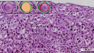
Cell cannibalism, or the formation of cell-in-cell structures, occurs when one cell becomes enclosed within another. This phenomenon is often seen in cancer and is thought to help tumour cells cope with harsh conditions such as lack of nutrients or exposure to chemotherapy and radiotherapy. In oral squamous cell carcinoma, the most common type of oral cancer, these structures have been linked to a worse prognosis.
The study investigates how and why these cell-in-cell structures form, and what they reveal about tumour behaviour and treatment resistance.
Professor Dan Lambert, Deputy Dean of the School of Clinical Dentistry said: “Our main goal is to find out how the tumour microenvironment (the surrounding cells and molecules that support cancer growth) influences this process.
“We’re particularly interested in how cancer-associated fibroblasts, a key component of that environment, might be helping to drive cell cannibalism.”
To address these questions, the researchers combined several innovative approaches. Analysing public cancer databases, they identified gene expression patterns linked to oral cancer cases. In the laboratory, they recreated the tumour environment using three-dimensional ‘spheroid’ models, which allows researchers to observe how different cell types interact in conditions that closely resemble real tumours. They also used spatial transcriptomics, a cutting-edge technology that maps gene activity within tissue samples, to see exactly where and how these structures form.
Findings show that the number of cell-in-cell structures found in the tumour spheroids are increased in the presence of cancer-associated fibroblasts. Areas rich in these structures also display gene expression patterns associated with higher metabolic activity, suggesting that the cells inside are working harder to survive. The team is now investigating whether tiny particles released by fibroblasts, known as extracellular vesicles, play a role in triggering this phenomenon.
For clinicians, this research could pave the way for recognising cell-in-cell structures as a new prognostic marker. As these structures are already visible in routine pathology slides, identifying them may help doctors better predict how a cancer might behave. For patients, the hope is that this knowledge will ultimately lead to more personalised treatments that target the survival mechanisms of tumour cells.
Leonardo de Oliveira Siquara da Rocha, Postdoctoral Researcher added: “This work gives us a completely new perspective on how cancer cells interact with their surroundings. By understanding the biology of cell cannibalism, we may be able to identify new ways to stop tumours from growing or becoming resistant to therapy.”
Clarissa Gurgel, Professor at the Federal University of Bahia's School of Dentistry and Vice-Director at the Gonçalo Moniz Institute, added: “By engulfing neighbouring cells, cancer cells gain metabolic support, enhance clonal selection, and promote immune evasion and invasion. In addition, unravelling the mechanisms underlying tumour cell cannibalism may also inspire therapeutic strategies aimed at inhibiting pathways associated with acidosis, among others.”
The project is part of a long-standing collaboration between the University of Sheffield and Fiocruz-Bahia, supported by the Brazilian National Council for Scientific and Technological Development (CNPq). In 2021, it was selected for the Newton Advanced Fellowship Grant, a prestigious programme that promotes research partnerships between the UK and overseas institutions.
Looking ahead, the researchers hope their findings could help integrate cell-in-cell detection into standard cancer diagnostics and perhaps one day support the development of AI-based pathology tools that use these structures as an additional measure for prognosis and treatment planning.