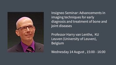
Insigneo Seminar: Advancements in imaging techniques for early diagnosis & treatment of bone disease

Event details
This event has taken place.
Description
We are pleased to welcome Harry van Lenthe a Full Professor of Biomechanics at the Biomechanics Section in the Department of Mechanical Engineering at KU Leuven (University of Leuven), Leuven, Belgium to give an Insigneo Seminar on 'Advancements in imaging techniques for early diagnosis and treatment of bone and joint diseases' on Wednesday 14 August 2024.
Brief Biography:
His research focuses on the quality of bone, and how it is changing during growth and aging. In addition, he is studying the interaction between bone and implants. He has been developing quantitative non-invasive methodologies, using state-of-the-art bioimaging, visualization techniques, and computational analyses, to assess bone quality. Main areas of expertise are micro-CT scanning and subsequent quantitative analyses. A special focus is on micro-CT-based finite element modeling using high-performance computing.
Harry van Lenthe has received several awards including the European Society of Biomechanics (ESB) Research Award (2000). He contributed to the scientific community amongst others by serving on the ESB council for several years before leading it as its president from 2020–2022. Since 2021 he is heading the Biomechanics Section at KU Leuven.
Title and Abstract:
Advancements in imaging techniques for early diagnosis and treatment of bone and joint diseases
Bone and joint health is of paramount importance to our wellbeing. Diseased bone, such as in osteoporosis and bone cancer, reduce the bone strength and increase the risk of fracture; diseased joints, such as in osteoarthritis, are associated with many functional restrictions, mainly due to pain. Both have negative impact on emotional well-being, and strongly reduce the quality of life of patients.
In this seminar, I will present novel imaging techniques that can facilitate earlier diagnosis of bone and joint diseases, which is crucial for providing better treatment to patients. Specifically, I will demonstrate the capabilities of the recently introduced photon-counting computed tomography for quantifying bone microstructure in vivo. I will share data from several anatomical sites and illustrate how image-based finite element models can help optimize treatment for patients suffering from bone metastases.
Location
53.382657550565, -1.47774535
When focused, use the arrow keys to pain, and the + and - keys to zoom in/out.

