
Equipment
Our multi-million pound facility houses multiple instruments for biological and soft-matter electron microscopy, based in custom-built laboratories, alongside office spaces.

Central to the facility is our Arctica cryo-electron microscope, which incorporates state-of-the-art automation and detection technology to determine the three-dimensional structure of biological molecules and cells.
More than 350 users from across the University of Sheffield currently access our equipment, which includes instruments that are generally found only at large pharmaceutical companies. This opens up the potential for research collaborations with small- to medium-sized businesses.
The standard access charge for EMF is £90 per hour. This includes basic consumables. However, if you need a specialised consumable then you may need to procure that separately. The charges may also be lower or higher depending on the type of funding, the techniques you need to use, the level of experience of the user. Please contact us (link) with more details of your work for a quote specific to your project.
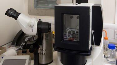
Tecnai Arctica cryo-electron microscope
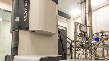
Ava is a 200 kV TEM with capabilities for room temperature and cryo conditions. This microscope is equipped with a 12 sample autoloader allowing for rapid loading and unloading of samples and a Volta phase plate for added contrast. The images from this microscope are recorded on a highly sensitive Falcon III direct electron detector or on a CETA detector, which is less sensitive but more robust. For automated image collection for SPA and tomography EMF uses EPU and FEI Tomography softwares respectively. This state-of-the-art cryoTEM is capable of producing sub 3Å structures in-house. All the cryoEM experiments at EMF are performed on this microscope.
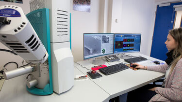
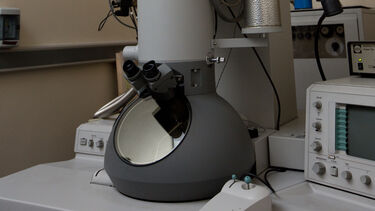
JEOL JEM-120i (“Jemma”)
Jemma is a latest generation easy to use 120 kV TEM with room temperature capabilities and side-entry sample holder capable of loading four samples in one go. This enables rapid, high-quality imaging. All room temperature TEM at EMF are performed on this microscope.
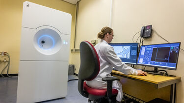
Scanning electron microscopes
Tescan Vega3 (“Vega”) LMU Scanning Electron Microscope with Oxford X-Max 50 EDS Detector - the Tescan Vega3 is a highly adaptable SEM instrument that operates in both high-vacuum and variable-pressure modes. It can work at multiple imaging modes delivering stable images with surface resolutions down to 3.0 nm. Its spacious vacuum chamber with 5-axis motorised eucentric stage is suitable for accommodating and effortlessly navigating large or bulky samples. Imaging of frozen samples is also an option.
The Oxford X-Max 50 is a sensitive elemental analysis detector. It excels at rapid detection of light elements in the microscopic sample. Its user‑friendly AZtec software provides real‑time mapping and reliable elemental identification.
All SEM imaging and sample elemental analysis is done on this instrument.
Transmission and scanning transmission electron microscopes
Quantity: 2
Plunge Freezers
EMF has two flash freezing robots -
- Leica cryo GP with temperature and humidity control, single-side face-on blotting for maximum control of blotting parameters. This instrument is great for SPA sample preparation, as well as ideal for blotting and freezing thick samples and cells for cryoET, due to the ‘back blotting’ capability.
- FEI Vitrobot Mach III with temperature control and angled blotting from both sides. Great for SPA sample preparation, creating a gradient of ice thicknesses for easier screening of optimal ice thickness.
Sectioning equipment
EMF has several ultramicrotomes for room temperature- and cryo-sectioning. Used to cut extremely thin slices of biological or soft materials—often just tens of nanometres thick—so they can be viewed clearly under the TEM.
Leica Cryo-CLEM system
This is an advanced research system that combines fluorescence (light) imaging with Cryo-EM-imaging to first locate features of interest using visible light, and then study their fine structure at much higher resolution in the EM.
Leica CPD
Used for gentle drying of sensitive SEM samples, while preserving their fine structure.
Computing capabilities
EMF maintains four GPU/CPU hybrid workstations with the latest image processing and 3D reconstruction softwares including Relion, Cryosparc, and Eman2.
Advice on data storage: While extremely powerful, SPA and cryo-tomography requires careful management of large quantities of data. A typical data collection session generates anywhere between 2 to 4 TB of data consisting mainly of cryoEM movie frames. In our experience another 6-8 TB storage space might be needed for data processing. While EMF provides computing capabilities for data processing no data storage space will be arranged by EMF. This is the responsibility of the individual users. For image processing on EMF workstations please arrange for external hard drives with at least 8 to 10 TB of storage space per dataset (this includes the storage for cryoEM movies). We do not advise storing cryoEM movies on external hard drives for long term storage.
Any data collected at EMF will be stored for 6 months. After that it is the responsibility of the individual user to transfer the data from EMF storage space to their own data storage arrangement. After 6 months all user data will be deleted.
The facility has been funded via the University of Sheffield’s Imagine: Imaging Life project, as well as grants from BBSRC, Wellcome Trust, Royal Society, and Wolfson Foundation.