Equipment
The Wolfson Light Microscopy Facility provides access to different types of imaging systems, enabling users to image their samples using multiple microscopy techniques.
This set of capabilities increases the potential for discoveries and maximises the amount of information that can be extracted from specimens.
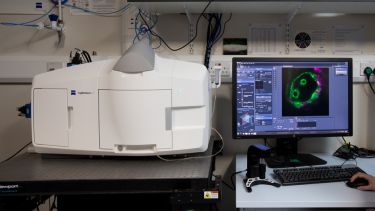
Confocal microscope
Optical sectioning of thick biological samples (<200uM) giving fantastic resolution at depth. Ideal for imaging tissues, developing systems and for profiling 3D structure. Our specialist systems also offer fast (spinning disk) as well as super-resolution (140nm) confocal microscopy (Airyscan confocal).
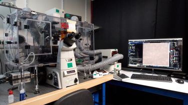
Widefield microscope
Camera-based microscopy systems that are ideal for the imaging of thin specimens like cultured cells and tissues. Our systems are equipped with the latest sCMOS and EMCCD cameras that are capable of detecting the faintest of signals at very fast speeds in both fixed and living cells.
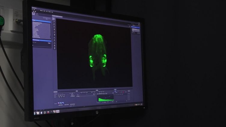
Lightsheet microscope
A novel imaging system for the gentle imaging of large specimens like developing systems or tissue spheroids.
SIM microscope
Super-resolution (120-140nm) imaging of thin samples (single cells).
STORM microscope
Super-resolution technique that can image fixed samples down to 20-50nm. This is great for studying the distribution and interaction of proteins and small molecules.
Stereomicroscope
For the screening of larger specimens and for the generation of extended focus images. We also have a Zeiss fluorescent Stereomicroscope.
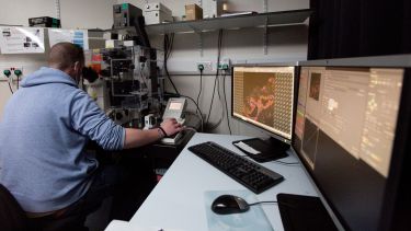
Image analysis suite
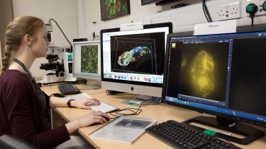
We have a suite of software for the analysis and presentation of multidimensional data, including Imaris, Arivis, Volocity and FIJI.

